Reproduction of David Maurice web site at Columbia | [ DavidMaurice.org home page ] |
SUMMARY:
Inadequacies of a standard technique of measuring aqueous flow are discussed
at different levels.
|
There are a number of techniques for measuring aqueous flow in humans by fluorometry, as described by Brubaker, who also gave a full discussion of sources of error. This revision of Brubaker's work will be based on the Protocol adopted by the European Community Network on Ocular Fluorometry (1990), since it appears to be a codification of current practice. In summary, this follows the Yablonski variation of Method 2 of Jones and Maurice, in which the total amount of fluorescein present in the anterior segment is estimated by adding that in the cornea to that in the aqueous, in each case multiplying the measured concentration by a volume factor. The time table of events is as follows. The measurements are taken with the Fluorotron Master® . |
0 hr Topical Fluorescein 4 hr
|
Modeling: assumptions incorrect. Data analysis: inefficient use. |
|
Instrumental errors have been well covered by Brubaker. The Fluorotron has a poor depth resolution and the true corneal concentration is estimated from the actual peak reading by an empirical function of the corneal thickness. |
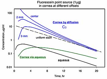
I suggest that a better approach would be to derive the
total amount of fluorescein in the cornea from the area under the corneal
concentration profile, as carried out in the tears by Joshi et al.
Deconvolution of the profile would seem to be another alternative, but I have no experience with it. |
|
|
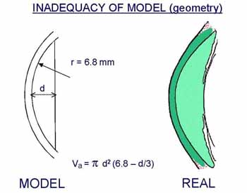
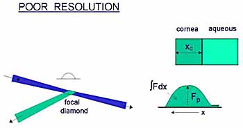
The importance of false assumption as to the geometry of the model depends on the purpose of the experiment, in particular, whether comparative or absolute flow values are required. |
|
The effect of exchanges with other compartments has been dealt
with by Brubaker, and how to correct for changes in diffusional loss across
the iris was explained by Anselmi et al.
Poor mixing in the anterior chamber is a nuisance that can be overcome by making eye movements. |
|
However, an unrecognized serious potential for error
is the uneven staining of the cornea.
Fluorescein can spread across the cornea by land or by sea: by diffusion through the corneal stroma or by passage in to and out of the well mixed aqueous. Rough calculations indicate that the sea route is very slow. |
|
Thus the principal mechanism of spreading is
by diffusion, and the distribution at any time can be readily calculated
and compared with the uniform staining required by the Yablonski method.
If fluorescein is applied as a spot near the corneal apex, values of central fluorescence are found at 4-7 hr. that can be an order of magnitude greater than assumed for uniformity. |
|
Of course, when a drop is instilled it does not penetrate
the cornea at one point only, but neither does it penetrate it uniformly.
Patches of deeper staining are often visible near the center.
There would seem to be a considerable risk of error from this source, therefore. |
|
A primary consideration in the procedure is the efficient
use of the patient and technician's time. It is probable that there is
little benefit to be gained by taking hourly readings. These could be concentrated
at the beginning and end of a period of preferably 4 + hour, leaving
the intervening stretch free for other activities.
In any case, my experience suggests that a one hour period is too short, in view of the spontaneous variations in aqueous humor fluorescein level in many subjects. This level drops an average of 25% in each hour, so that a +/- 2% inaccuracy in the aqueous, measurement, which can readily occur, translates into a +/- 8% error in the flow estimate. |
References: |
COMAC-BME Project on Ocular Fluorometry. Coimbra, Portugal. 1990. |
Anselmi P. Bron AJ. Maurice DM. Action of drugs on the aqueous flow in man measured by fluorophotometry. Exp. Eye Res. 1968. 7:486-96. |
Brubaker RF. The flow of aqueous humor in the human eye. Trans Amer. Ophthalmol. Soc. LXXX 1982. 391-474. |
Jones RF. Maurice DM. New methods of measuring the rate of aqueous flow in man with fluorescein. Exp. Eye Res. 1966. 5:208-220. |
Joshi A. Maurice DM. Paugh JR. A new method for determining corneal epithelial barrier to fluorescein in humans. Invest. Ophthalmol. Vis. Sci. 1996. 37:1008-1016. |
Yablonski ME. Zimmerman TJ. Waltman SR. Becker B. Exp. Eye Res. 1978. 27:135-142. |
Reproduction of David Maurice web site at Columbia | [ DavidMaurice.org home page ] |