 ;
;
In human, Bowman describes 60 lamellae by histology, but Pouliquen et al. 307 lamellae by TEM.

| SUMMARY: |
| The mammalian cornea differs from other connective tissues in three ways. |
| PROPERTY | INFERENCE |
|
TRANSPARENT SWELLS SHEARS |
Fine fibrils Fibrillar order: Interfibrillar forces. Interfibrillar forces. Not interwoven: Lamellar structure. Not interwoven: Lamellar structure. |
| Four puzzling features of the lamellar structure are discussed: |
|
| ; |
Slide 1. Cross-section of beef cornea observed under polarizing microscope. About 70 lamellae. |
|
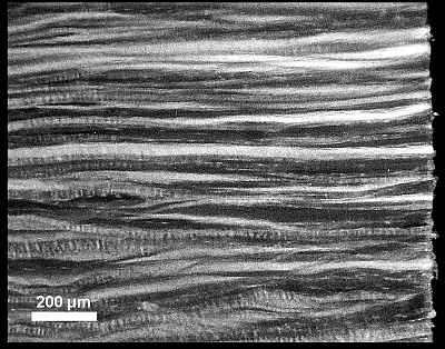
Slide 2. Low power TEM of beef stroma, showing 3-400 lamellae.
In human, Bowman describes 60 lamellae by histology, but Pouliquen et al. 307 lamellae by TEM. |
| |
Slide 3. Small piece of stroma lying flat at bottom of cuvette under saline, and observed by slit lamp. Slit vertical, i.e., perpendicular to plane of cornea. |
|
Slide 4. Striated appearance of very swollen stroma viewed in slit lamp as in Slide 3. |
 |
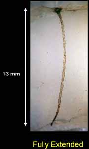
Slide 5. Showing how cohesive structures in stroma must be stretched to several times their length by swelling and even more by shear. |
 |
Slide 6. Method of marking stroma to demonstrate shear. |
 |
Slide 7.
Left, unstressed. Right, maximal shear. |
 |
Slide 8.
Left, unstressed. Right, maximal shear. Tissue shows greater freedom of movement as hydration increases. |
 |
Slide 9.
Left, unstressed. Right, maximal shear. Tissue stiffening again near maximum hydration |
|
Slide 10. Thick cross-section of swollen stroma crushed perpendicular
to face of section, showing further (irreversible) extension of structure.
|
| REFERENCES: |
| Bowman |
| Pouliquen et al. |