Presented at the ARVO '98. Invest. Ophthalmol. Vis. Sci. (1998) 39, S1030.
INTRODUCTION
The long term changes in morphology of keratocytes are to be followed
under a confocal (Koester, slit-scanning) microscope by repeatedly relocating
a marked area of the mouse cornea.
This is facilitated by being able to rotate the animal around
the center point of the eyeball in a system of gimbals, so that the cornea
remains in focus as the area under observation is shifted.
CONSTRUCTION
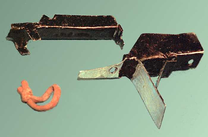
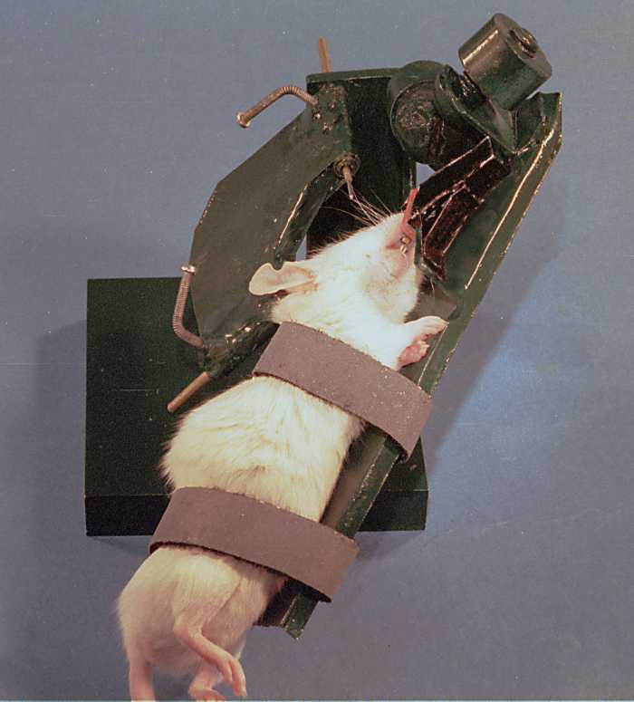
A device has been fabricated with hand tools from thin metal sheet
and telescopic brass tubing, joined by epoxy glue.
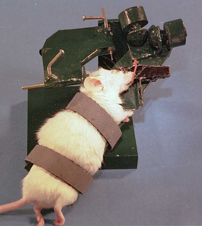
To reduce eye movements, the head is fixed in a clamp based on the
design of Erickson, except that the metal screw is replaced by a rubber
band.
The eye proptoses spontaneously in an anesthetized animal when the clamp
is fixed to the head.
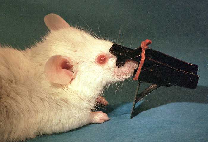
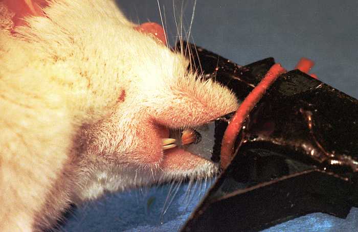
Trauma from the polished metal plate pressing on the palate is not observed.
OPERATION
The head-clamp is attached to a counterbalanced platform by means of
magnets so that its position can be adjusted. Velcro straps hold the mouse
on to the platform.
The center of the globe is moved to the center of rotation, guided by
retractable pointers sliding through the gimbals.
The platform can be rotated to bring most of the corneal surface into
the field of view of the microscope; it can be fixed in position by set
screws.
FUNCTION
Movement of the cornea from respiration and heartbeat is negligible
when the head clamp is properly applied and an applanating objective is
used.
A marker inserted in the stroma can be identified and brought into focus
vertically under the objective in a minute or two.