
D Maurice, S Velilla, anb L-S Chuang
Columbia University Department of Ophthalmology
presented at ARVO, 1999
| SUMMARY: |
| Usually, studies of the penetration of a drug from the tears to the
retina and vitreous are carried out in the rabbit, somewhat as follows:
Standard Technique
|
| ERRORS IN STANDARD TECHNIQUE |
|
1. Contamination 2. Penetration After Death |
|
|
| AVOIDANCE OF ERRORS |
|
In the present experiments we attempted to avoid these errors by: 1. Small subconjunctival injection 2. Freezing eye after death |
| EXPERIMENTAL PROCEDURES |
|
| TRACERS | |
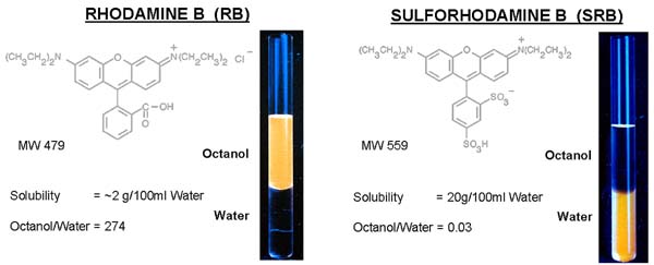
| Dyes were used as tracers because their distribution
after injection is visible. Fluorescent dyes can be determined in very
low concentrations.
Rhodamine B (RB) and Sulforhodamine B (RSB) were chosen for representing extremes of lipophilicity. |
|
|
|
| INJECTION | |
| A measured volume, ~ 0.2 µl, of 10% SRB or 1% RB injected RE through glass electrode near equator nasal to superior rectus. The solution was made viscous with hyaluronic acid to prevent regurgitation. |
 |
| FREEZING | |
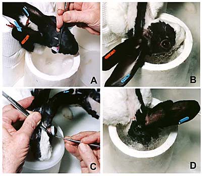 |
The animal is elevated on a platform.
After death, the right eye is proptosed (A) and dipped into liquid nitrogen (B) for about 1 min. The left eye is then proptosed (C) and both eyes plunged into liquid nitrogen (D) until boiling ceases. |
| PREPARATION OF CORE |
|
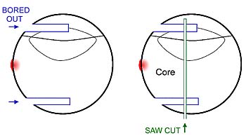
| To firmly anchor the frozen head a 1/4 inch hole is drilled through it
and a bolt is passed through it.
The bolt is fixed in a yoke and attached to the tool post of a hobbyists' lathe. A 16 mm diameter channel is bored into the globe with a chilled tool. The core is separated with a saw. |
 |
| SEPARATION OF TISSUES | |
| The core is lyophilized for 24 hr.
The vitreous can be lifted out cleanly in one piece. The retina comes off in flakes on pressing with a tool. The RPE sometimes comes with it, sometimes not. The choroid is scraped off the sclera. A colored spot is seen centrally on the inside of the sclera of injected eyes. It becomes spread out in the longer term experiments. The sclera can be split form conjunctival tissue. The fluorescent label is extracted from the tissue samples and measured in a fluorometer that can estimate 1 picogram. |
 |
| RESULTS |
|
|
Rhodamine B
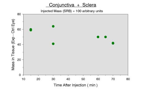
Conjunctiva + Sclera |

|
|
Rhodamine B
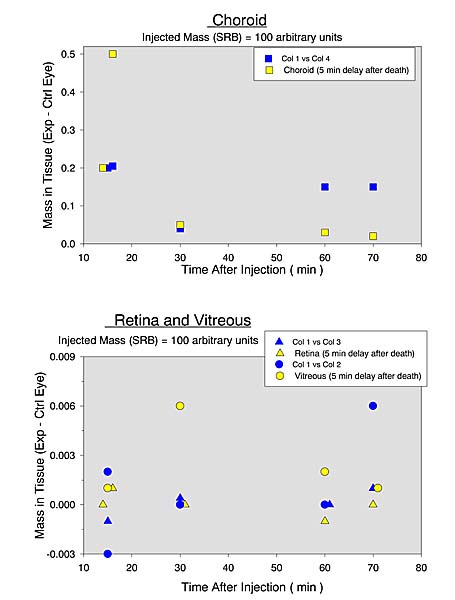
Choroid Rhodamine B Retina and Vitreous |

|
| Sulforhodamine B Conjunctiva + Sclera |
|
|
Sulforhodamine B
Choroid Sulforhodamine B Retina and Vitreous |
|
| DISCUSSION |
|
| CONCLUSIONS |
|
| REFERENCES: |